These ipRGCs are a morphologically and physiologically heterogeneous population that project widely throughout the brain and mediate a wide array of visual functions ranging from photoentrainment of our circadian rhythms. Recent works have identified and characterized several anatomically and functionally distinct ipRGC subtypes and have added strong.

One class of these cells the midget retinal ganglion cells mRGC are the most numerous.
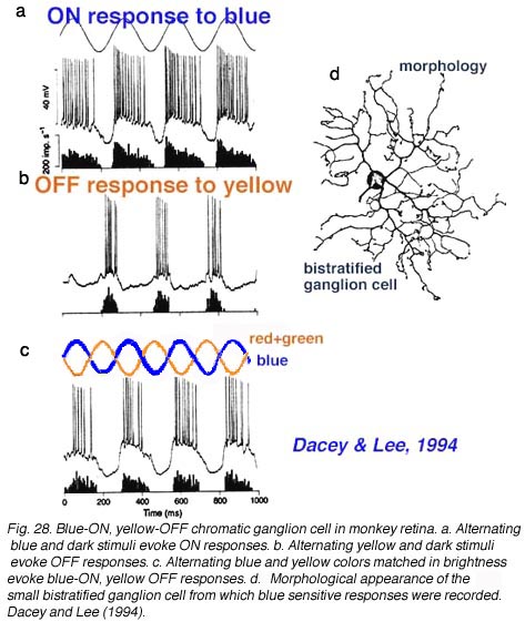
Retinal ganglion cells function. Ganglion cells are the output retinal neurons that convey visual information to the brain. There are 20 different types of ganglion cells each encoding a specific aspect of the visual scene as spatial and temporal contrast orientation direction of movement presence of looming stimuli. Ganglion cell functioning depends on the intrinsic.
Ganglion cells function Distribution of retinal ganglion cells ganglion cells function. Retinal ganglion cells function RGC encode various features of the visual environment and. Distribution of retinal ganglion cells.
Retinal ganglion cells function is relatively evenly distributed in the. Retinal ganglion cells. Development function and disease.
Baehr W Wensel T Hageman G Wu S Zack D. 21251506 Indexed for MEDLINE Publication Types. Retinal Diseasesphysiopathology Retinal Ganglion Cellscytology.
Retinal Ganglion Cellsphysiology Grant support. The function of retinal ganglion cells RGCs can be non-invasively assessed in experimental and genetic models of glaucoma by means of variants of the ERG technique that emphasize the activity of inner retina neurons. The best understood technique is the Pattern Electroretinogram PERG in response to contrast-reversing gratings or checkerboards which selectively depends on the presence.
In the vertebrate visual system all output of the retina is carried by retinal ganglion cells. Each type encodes distinct visual features in parallel for transmission to the brain. Various interneurons by the retinal ganglion cells RGC.
These are the output cells of the human eye and consequently their properties limit the signal that travels to the rest of the brain. One class of these cells the midget retinal ganglion cells mRGC are the most numerous. Near the fovea they appear to sample a single.
The function of retinal ganglion cells RGCs can be non-invasively assessed in experimental and genetic models of glaucoma by means of variants of the ERG technique that emphasize the activity of inner retina neurons. The best understood technique is the Pattern Electroretinogram PERG in response to contrast-reversing gratings or checkerboards which. Retinal ganglion cells RGCs are the projection neurons of the retina and transmit visual information to postsynaptic targets in the brain.
While this function is shared among nearly all RGCs this class of cell is remarkably diverse comprised of multiple subtypes. The discovery of a new class of intrinsically photosensitive retinal ganglion cells ipRGCs revealed their superior role for various nonvisual biological functions including the pupil light reflex and circadian photoentrainment. Recent works have identified and characterized several anatomically and functionally distinct ipRGC subtypes and have added strong.
Beginning at the first synapse in the retina lateral inhibition shapes signals creating antagonism between local center and global surround regions of visual space. Retinal ganglion cells RGCs were the first visual neurons to be described in terms of center-surround receptive fields 1. Where Are Retinal Ganglion Cells 17 mei 2021 Pin On Retina.
Pin On Trb. Sight The Sense Of Vision Medical Terminology Study Human Anatomy And Physiology Medical Anatomy. For More On Retina Histology And To See How This Diagram Maps Onto Histological S Basic Anatomy And Physiology Human Anatomy And Physiology Optometry Education.
How The Retina Works Detailed. The retina communicates visual information to the brain through the action potentials of retinal ganglion cells RGCs. This population of neurons consists of more than thirty distinct types each of which covers the retina to reliably encode its part of the visual message 1 2.
Retinal ganglion cells process visual information that begins as light entering the eye and transmit it to the brain via their axons which are long fibers that make up the optic nerve. There are over a million retinal ganglion cells in the human retina and. The melanopsin-expressing intrinsically photosensitive retinal ganglion cells ipRGCs are a relatively recently discovered class of atypical ganglion cell photoreceptor.
These ipRGCs are a morphologically and physiologically heterogeneous population that project widely throughout the brain and mediate a wide array of visual functions ranging from photoentrainment of our circadian rhythms. Discovery and function of ipRGCs The retina is a highly organized structure where the cell bodies of distinct neuronal types reside in well-defined nuclear layers and make synaptic connections in two distinct plexiform layers Figure 1. In the visual system a group of neurons called bipolar cells collects light-evoked photoreceptor signals in the outer retina and relays these signals to the inner retinal ganglion cells.
From there the images are projected as information to the brain. There are at least 14 bipolar cell types and the unique way they transform the photoreceptor signals allows the inner retinas neural circuits to form a visual description of the world.