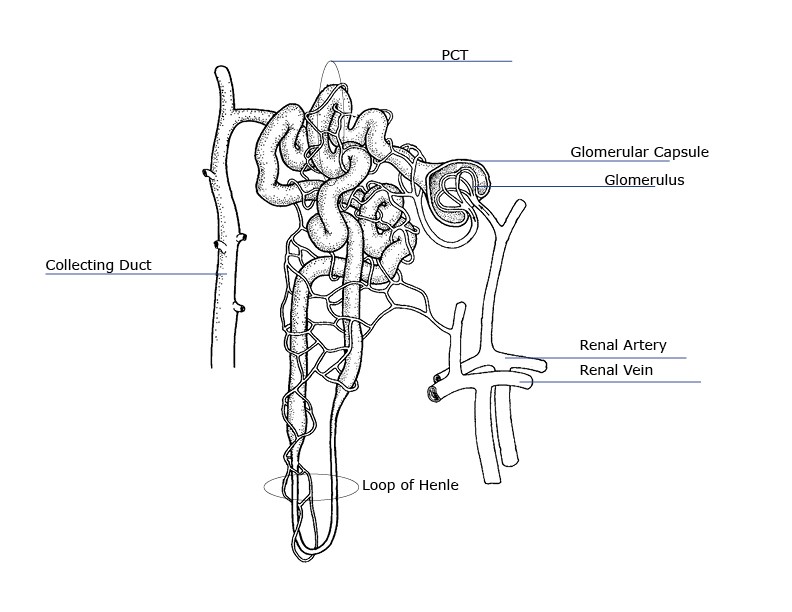
A nephron is the unit of structure and function in the kidney. This is an online quiz called Label a Nephron.

And help to sieve or filter wastes or toxins materials from the blood and expel them outside the human body.
Nephron diagram to label. The nephron is the microscopic structural and functional unit of the kidney. It is composed of a Diagram left of a long juxtamedullary nephron and right of a short cortical nephron. Proximal convoluted tubule lies in cortex and lined by simple cuboidal epithelium with brush borders which help to increase the area of.
Site at which most of the tubular reabsorption occurs. Between the loop of Henle and the collecting duct. Selective reabsorption and secretion occur here.
Descending loop of henle. Ascending loop of henle. The first slide is an overview of the urinary system that shows the kidneys ureters urinary bladder and urethra.
Students drag labels to the structures on the slide. Also the diagram shows the relationship between the aorta vena cava and the renal vessels. While these arent part of the urinary system they are important in the physiology of the kidney.
Diagram A Nephron Diagram Kidney Elegant Internal Structure labeled diagram of a nephron and its location and functions most of the nephron lies in the outer region of the kidney the renal cortex ly one part the loop of henle enters the central part of the kidney the renal medulla Kidney Cross Section Anatomy Diagram human body health. This is an online quiz called Label a Nephron. There is a printable worksheet available for download here so you can take the quiz with pen and paper.
From the quiz author. Printable kidney and nephron anatomy labeling Human body unit study Anatomy and physiology Biology lessons. Feb 1 2018 - This shows a kidney and the nephron with arrows that point to specific structures within the kidney for students of anatomy to label.
This is an online quiz called Label a Nephron. There is a printable worksheet available for download here so you can take the quiz with pen and paper. A nephron is the unit of structure and function in the kidney.
Each nephron is a coiled tube held together by a tough fibrous connective tissue. In humans a healthy adult has 1 to 15 million nephrons in each kidney functioning together to filter blood from all its impurities. They also regulate blood pressure control electrolytes and regulate blood pH.
Label This Diagram Of A Nephron. Find this Pin and more on Study Timeby Aurora Kay. Answer to Label this diagram of a nephron.
A nephron is the functional component of the kidneys structure. It is vitally utilized for the detachment of water ions and small molecules from the blood. And help to sieve or filter wastes or toxins materials from the blood and expel them outside the human body.
It also helps to the reappearance of desirable molecules back into the blood. Its main function is completed by ultrafiltration. This is how you can draw the diagram of Nephron step by step.
Please dont forget to subscribe my channel for more videos like this. On the diagram below label the major parts of the kidney. Fill in the blanks below to trace the flow of fluid through a cortical nephron.
The first line. Discover best Nephron Diagram images and ideas on Bing. Updated Nephron Urinary System Diagram.
Blank Diagrams Blank Kidney Nephron Diagram. How to draw step by step nephron diagram with double pencil method step by step for class 10 student by using some new tricks which helps to draw the structu. Label the structures in the nephron diagram below see diagram.
Proximal convoluted tubule Glomerulus. Click on download link below to download the match-each-lettered-structure-in-the-diagram-of-the-nephron PDF for free solar cell structure diagram PDF results Download PDF solar-cell-structure-diagram for free at This Site. Nephron Diagram on Final ie.
Renal Corpuscle Proximal Convoluted Tubule Distal Convoluted Tubule Collecting Duct Loop of Henle Terms in this set 34 Renal Corpuscle. Consists of glomerulus capillaries enclosed within Bowmans capsule. Outer parietal layer is composed of simple squamous epithelium.
Inner visceral layer consists of podocytes that wrap. For students in Biology 1002 try this technique on the following diagram of a nephron. Unfortunately they find out on the lab exam that they wont be asked to simply label a reference diagram that they have studied but they are asked to draw a diagram of a specimen and label it.
Students that have not developed the interpretive skills cannot effectively draw and label their own diagrams. How to draw nephron in easily- 10 cbse. Best way to draw nephron easily.